|
|
Post by kungfuzu on Dec 16, 2022 20:40:23 GMT -8
|
|
Brad Nelson
Administrator
עַבְדְּךָ֔ אֶת־ הַתְּשׁוּעָ֥ה הַגְּדֹלָ֖ה הַזֹּ֑את
Posts: 11,047
|
Post by Brad Nelson on Dec 17, 2022 11:03:55 GMT -8
And there would be extra credit just for spelling his name right. I grabbed some more moss outside and then found many of these: Voticella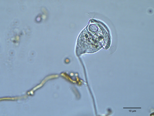 The above image is from the web but that's about exactly the resolution that I saw in my scope. Let me tell you, the micro world is creepy...creepier than Joe Biden around children. Well, maybe not that bad. But I saw one of these with its stalk attached to a piece of moss (and it was surrounded by a whole colony of other Voticella). And it appeared to be in the middle of presumably mitosis. It was duplicating itself. Or was I just seeing another Voticella behind it and the appearance of the cell beginning to pinch in the middle was just an optical illusion? So I sat and watched it under the microscope for about 30 minutes. My eyes were watering from the strain after that time. But this was indeed a Voticella (a single-celled Protozoa) that was in the middle of duplicating itself. When I first saw the creature, it was fairly round and didn't have the mouth parts that are opposite to the stalk. It took a fair while, but finally the cells separated. But before they were fully separated, you could clearly see that both of these once-rounded creatures were quickly developed the mouth parts...even as you watched. Maybe in 4 minutes the cells were no longer round but more oblong and with the mouth parts. I had no idea it could happen that fast. But then what do I know? Very little...which is one reason I got the scope. But the new Vorticella stayed connected to its parent for another 15 minutes or so. I watched and watched figuring that something must happen. It would sprout its own stalk or something. I had glanced in and out of the eyepieces because your eyes really do need some relief after a while. And one of the times after glancing back in, I just caught the newborn skedaddling off on its own at such a speed that I had no chance of trying to follow it. And, of course, I've since read on that Wiki page that they have a free-swimming stage before they attach themselves to something with a stalk. No doubt this at least temporarily homeless Vorticella had gone looking for a cardboard box. And, of course: |
|
Brad Nelson
Administrator
עַבְדְּךָ֔ אֶת־ הַתְּשׁוּעָ֥ה הַגְּדֹלָ֖ה הַזֹּ֑את
Posts: 11,047
|
Post by Brad Nelson on Dec 19, 2022 11:55:03 GMT -8
Here's a one-minute video of some creature I found in the sample that I took from Kitsap Lake. This is only the second video I've ever uploaded so I'm new to this.
I had Christmas music (by coincidence...this is not a production value) going on in the background so that got captured with the video. YouTube has very aggressive copyright restrictionw so expect this sound to be automatically removed at some point...or for the entire video to get nixed. I'm not sure what will happen.
The quality of the video is degraded from the original. I'm not sure what the magic is so that you can upload without a significant loss of detail.
It actually looks pretty good on my iPad Pro via the Safari web browser. But in either the YouTube app on my iPad, or on YourTube on my Windows 10 machine in FireFox, it looks a lot blurrier. I don't know why that is.
Okay...I changed the quality setting manually to 1080p (that is the resolution that it was filmed in) and that seemed to fix it in the FireFox web browser in Windows 10. But it didn't do anything for the YouTube app on the iPad...and app that has always been a little flaky.
---
Yeah, YouTube's AI picked that up right away as a copyrighted tune. Because this is not a commercialized video, it meant no restrictions. But I used their tools to replace it with a Christmas tune. They had a convenient list of non-copyrighted tunes to choose from. And the soundtrack was swapped using the YouTube Studio tools.
|
|
Brad Nelson
Administrator
עַבְדְּךָ֔ אֶת־ הַתְּשׁוּעָ֥ה הַגְּדֹלָ֖ה הַזֹּ֑את
Posts: 11,047
|
Post by Brad Nelson on Dec 19, 2022 14:01:28 GMT -8
Here's a still shot of the above: 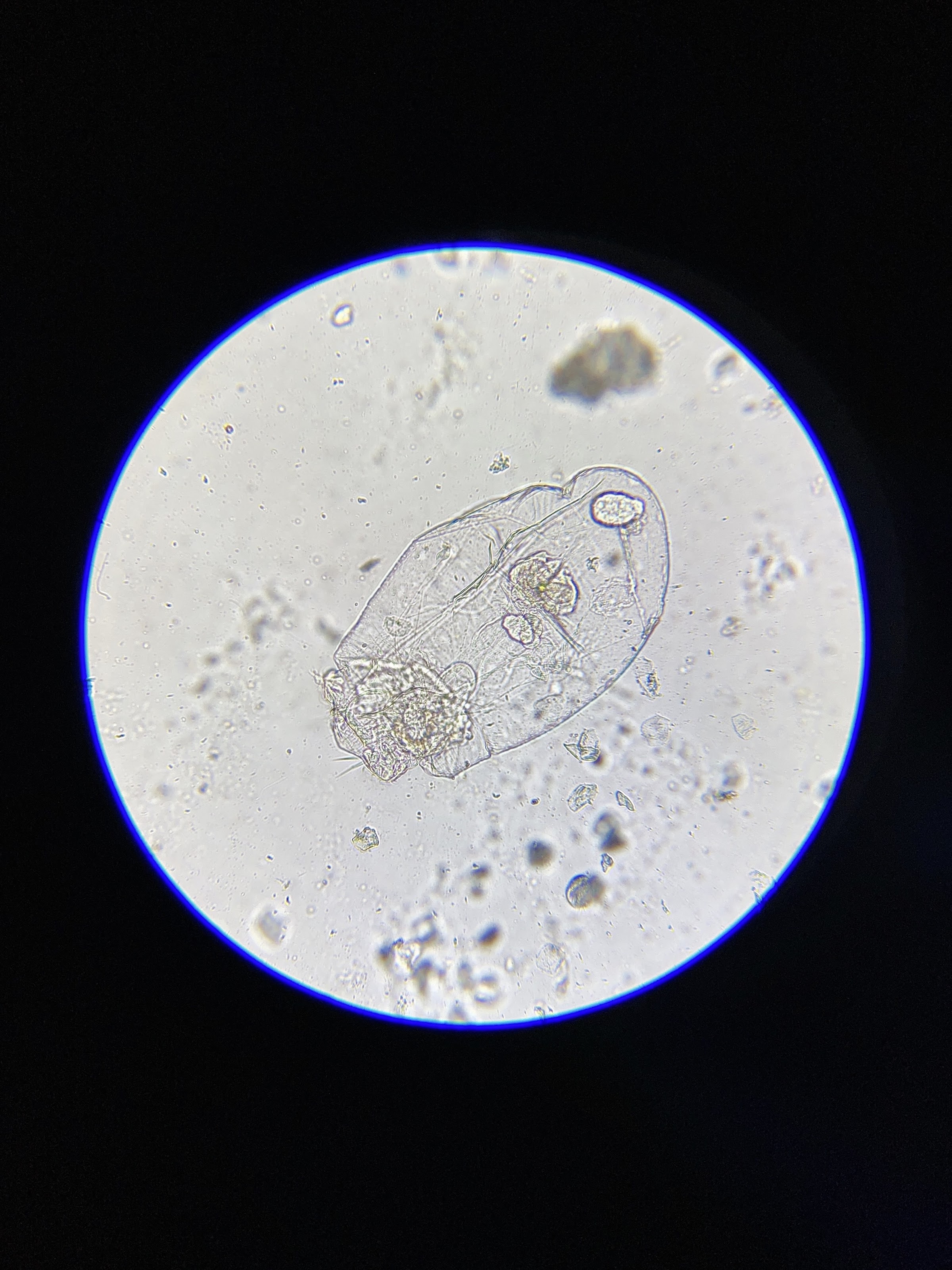 Larger View Larger ViewBoth the above photo and the video are uncropped. It's interesting that the still photo is cropped less (compared to what you can view through the microscope eyepieces) than the video. I don't know why. There's a rhyme and reason to all of this. But it is what it is. I could, of course, for presentation purposes crop either or both, turning them 90 degrees in the process (especially suited for the video) if desired. |
|
Brad Nelson
Administrator
עַבְדְּךָ֔ אֶת־ הַתְּשׁוּעָ֥ה הַגְּדֹלָ֖ה הַזֹּ֑את
Posts: 11,047
|
Post by Brad Nelson on Dec 19, 2022 15:21:47 GMT -8
Bought a pair of these rubber eyecups. It definitely improved things. Although the "eye relief" (how close you have to be to the eyepieces) is small, these work if you roll the rubber back around the edges...which is what these are made for, or at least another way they can be used. Having your eyes against rubber is more comfortable than against metal.
|
|
Brad Nelson
Administrator
עַבְדְּךָ֔ אֶת־ הַתְּשׁוּעָ֥ה הַגְּדֹלָ֖ה הַזֹּ֑את
Posts: 11,047
|
Post by Brad Nelson on Dec 19, 2022 21:33:25 GMT -8
Just taking a second attempt at photographing the prepared slide of the paramecium. 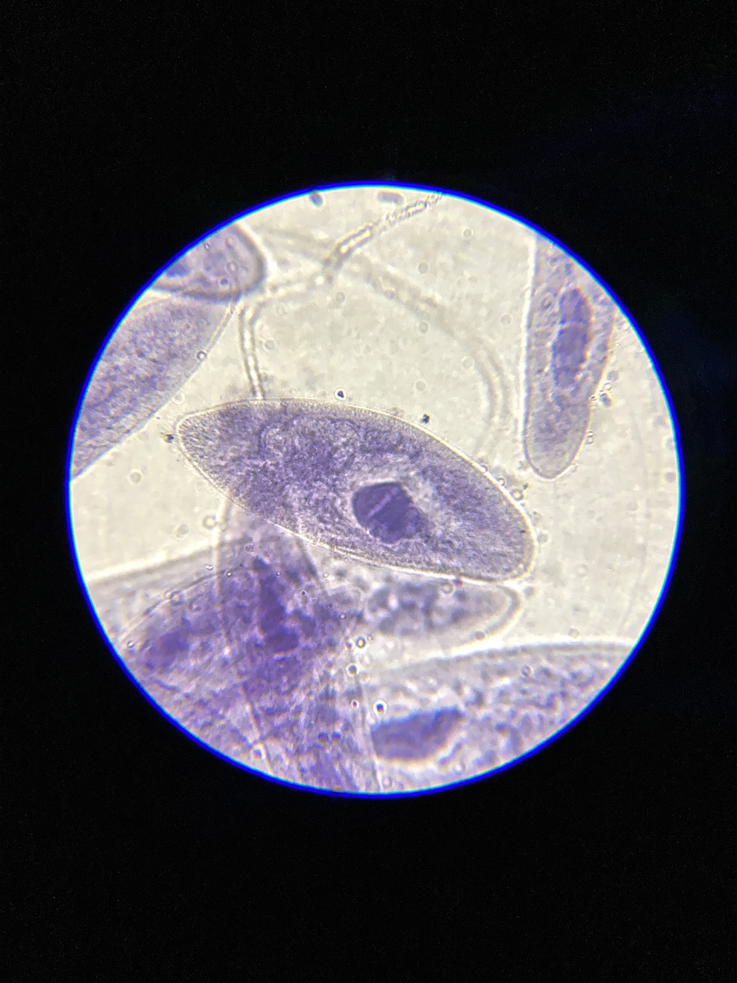 Larger View Larger View |
|
Brad Nelson
Administrator
עַבְדְּךָ֔ אֶת־ הַתְּשׁוּעָ֥ה הַגְּדֹלָ֖ה הַזֹּ֑את
Posts: 11,047
|
Post by Brad Nelson on Dec 20, 2022 15:32:35 GMT -8
Always working on my photo editing skills. This is the same photo as above but edited with Affinity Photo on my iMac.  Larger Version Larger VersionThis one uses a curves adjustment (for contrast), a little white balance adjustment (so the whites are a little less yellow), and uses a high pass filter trick I once found online as a way to do sharpening. All of these are in non-destructive “adjustment” layers. I think this edit is a little less heavy-handed than the previous one. Granted, these are somewhat subtle differences. |
|
Brad Nelson
Administrator
עַבְדְּךָ֔ אֶת־ הַתְּשׁוּעָ֥ה הַגְּדֹלָ֖ה הַזֹּ֑את
Posts: 11,047
|
Post by Brad Nelson on Dec 22, 2022 15:41:17 GMT -8
Still playing around with a workflow for photos. Below is a 1000x shot (using oil on the 100x oil lens and using the 10x ocular eyepieces). It's a commercial slide of frog blood.
Okay, I over-saturated the red a little bit. But the workflow that I'm finding is easy and works well is:
1) Shoot on the iPhone SE II with the microscope adapter through the left eyepiece (on the binocular scope). I can still view the slide through the right eyepiece easily enough. I use the ProCamera app on the iPhone and set it to manual focus. It would be difficult to shoot anything with a phone if the phone is controlling focus. What I do via the ProCamera app is zoom to 200% (it's a digital zoom), make any fine-focus adjustments on the fine focus knob of the microscope (while viewing the phone's screen through a magnifying glass), and then zoom back out to 100% and take the shot via a delay timer to avoid any camera shake. 2) Transfer photo to the iPad Pro via bluetooth (wireless and easy). 3) Edit using the built-in editing tools in the default Apple Photos app. I first adjust the "Definition" … a feature analogous to the "Texture" and/or "Clarity" of other software. I then up the contrast a little, sharpen a little, and up the vibrance (a special form of saturation) a little (a little too much in this case). Then I upload it to Google Photos via the Google Photos app. I can fuss in the more expensive photo apps (Photoshop, Affinity Photo, Pixelmator Photo), but I can't honestly say that for this use they do any better. And it takes a lot more work with those apps, if only because of the extensive importing and exporting needed via Apple's convoluted and hard-to-use file system. 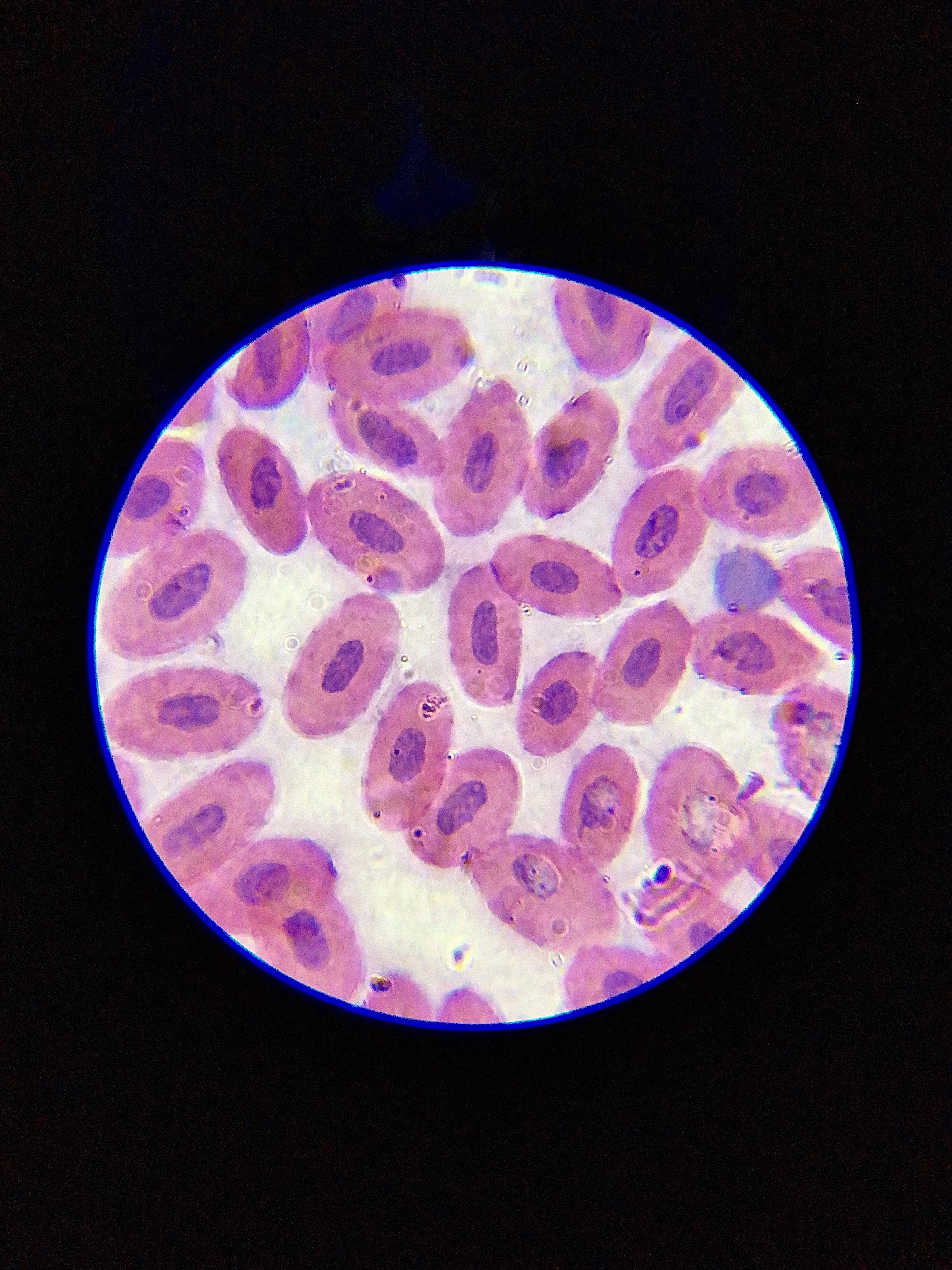 Larger View Larger View |
|
|
|
Post by kungfuzu on Dec 23, 2022 6:15:15 GMT -8
This brings up a question which I have been pondering regarding the various photos. With all the software usage, modifications, etc..... are we seeing the real pictures or is what we are seeing the equivalent of some of those NASA photos of deep space to which some artists add color in order to interest the general public?
I understand playing with film has always been part of photography, but I am interested in at what point editing becomes invention.
|
|
Brad Nelson
Administrator
עַבְדְּךָ֔ אֶת־ הַתְּשׁוּעָ֥ה הַגְּדֹלָ֖ה הַזֹּ֑את
Posts: 11,047
|
Post by Brad Nelson on Dec 23, 2022 7:27:46 GMT -8
A pretty rich red or blue stain (typically Methylene Blue) is common. And those stains are anything but subtle. Aside from the green of algae (either the various plants that use chloroplasts for photosynthesis or the animals that ingest algae and then have green inside them), it seems most of this microscopic life is semi-transparent...and certainly mostly colorless. That's where staining comes in as well as various colored photo filters ( Rheinberg filters) and other lighting techniques. I think you can use some stain on live specimens but, of course, it works very well when they are dead. Staining and other techniques are used in order to observe structure. But the philosophical issue is that this is a world where eyes like ours don't really exist. Much like deep sea creatures that you see in documentaries which are almost all transparent, color doesn't come into the equation in the world they inhabit. But particularly for scientific, medical, or even hobby or educational use, being able to see more structure inside these plants and animals is useful or interesting. There's also the artistic or presentation aspect. The Rheinberg filters are useful just to make the photos look a little more interesting than the typical back-lit washed-out light shades-of-gray on a bleached white background. And I would suppose that if the photos are labeled as such (and they sometimes are), there's no harm, no foul. I think with this microscopic stuff, it's an inherent artifice, as it were, just to be able to see them at all. We have to take these critters out of their natural environment, put them under a glass slide, and use a microscope. But since all that most of these creatures do is just swim around in water or walk around on moss, it's probably not all that unnatural. My beef is mostly with landscape photography where it has become the norm to take a photo of nature through what you could call an impartial lens and then subject it to an acid-trip of photo effects and manipulation on the back end. What you have then is no longer even marginally real. And, to my eye, these photos are garish, ugly, and do a disservice to nature:  To me, such photos show a lack of good taste. And, believe me, I've read deep inside of what is going on. Nature is not held in esteem. It is something to be blotted-out and redrawn. How often I've heard or seen some idiot professional photographer talk about removing little bits of this or that from a scene because it is "distracting" to what their vision of the photo should be. No, it's a photo, you idiot. Unless you are removing things because you are a commercial photographer (where presentation of a perfect image has at least the reason of money behind it), then you are just a technical hack. You are bending nature to your will. And your only guiding influence seems to be Playskool where lots of bright, shiny primary colors are the only things that matter. Granted, a certain amount of editing is understandably and reasonable. In many cases, your eye can see much more than can be recorded by a camera. So you might need to get more detail from the shadow and highlight areas, bump the contrast, etc. And there is nothing more basic to a photograph than the framing...deciding where to crop it. That's artificial, of course, but inherent to the format. But this trend toward psychedelia is clearly the result of the Snowflake culture having reached deep down into the arts. God forbid we might (even in just some esoteric fashion) admire God's handiwork, something above us and that we didn't create. But, no, these hack "artists" must put their stamp on it and thereby create little more than graffiti. But as for the microscope work I've seen, this Playskool approach has yet to infect it to any significant degree. |
|
|
|
Post by kungfuzu on Dec 23, 2022 13:57:07 GMT -8
Couldn't agree more!
|
|
Brad Nelson
Administrator
עַבְדְּךָ֔ אֶת־ הַתְּשׁוּעָ֥ה הַגְּדֹלָ֖ה הַזֹּ֑את
Posts: 11,047
|
Post by Brad Nelson on Dec 24, 2022 23:11:31 GMT -8
This next shot is a result of “focus stacking”. I took five photos of the same fixed subject, each with a different focus point (theoretically starting in the background and ending in the foreground). And it worked pretty well. The subject is rhizopus, whatever that is. Well, yes, it is exactly as it looks. It is a form of fungi. I found the focus stacking worked better (and was easier and faster) in Affinity Photo than it was in Photoshop.  Larger View Larger View |
|
Brad Nelson
Administrator
עַבְדְּךָ֔ אֶת־ הַתְּשׁוּעָ֥ה הַגְּדֹלָ֖ה הַזֹּ֑את
Posts: 11,047
|
Post by Brad Nelson on Jan 3, 2023 14:09:42 GMT -8
Just some moss. But I thought it looked cool. 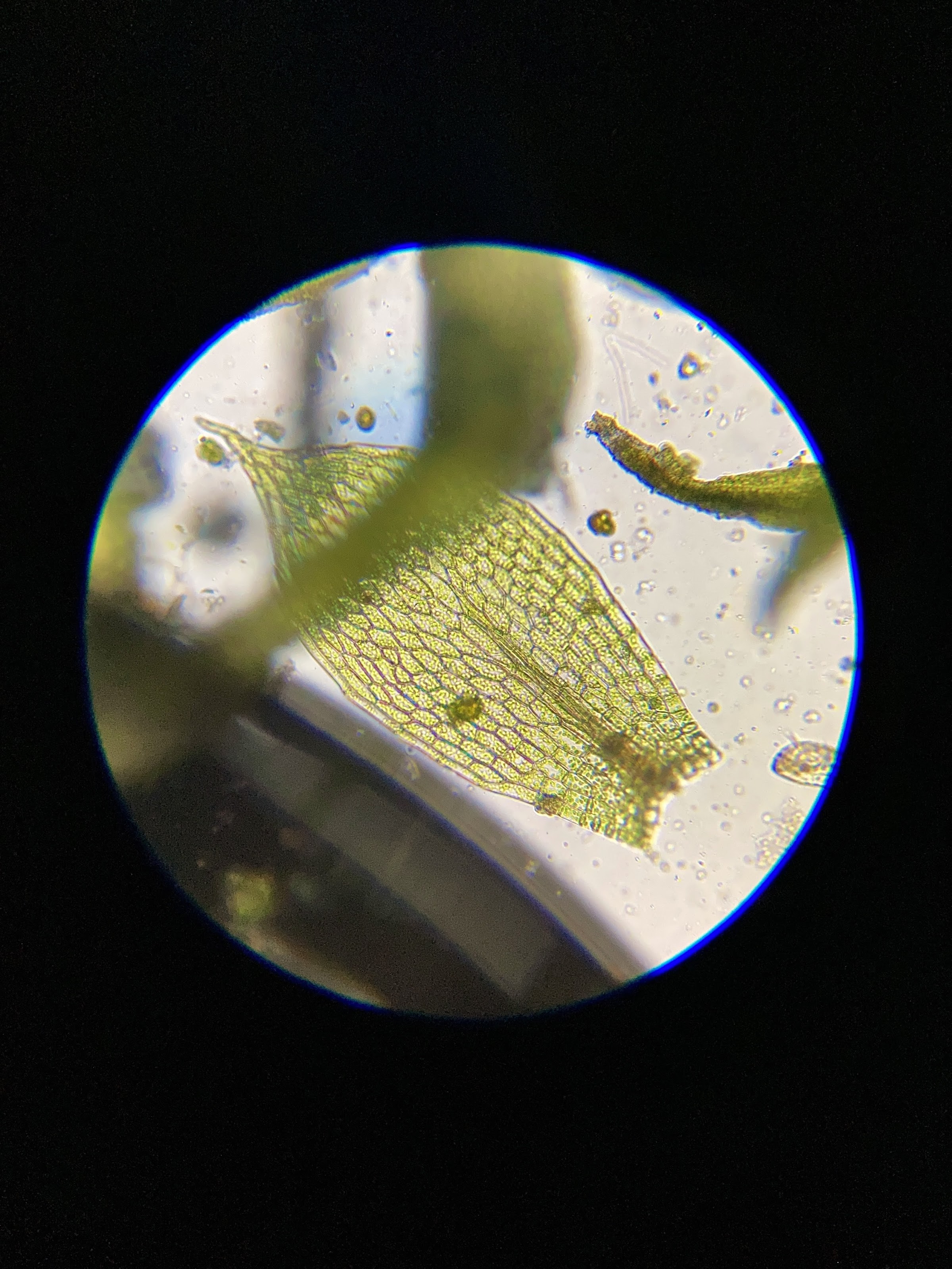 Larger View Larger View |
|
|
|
Post by kungfuzu on Jan 3, 2023 15:07:28 GMT -8
That reminds me of this.  |
|
Brad Nelson
Administrator
עַבְדְּךָ֔ אֶת־ הַתְּשׁוּעָ֥ה הַגְּדֹלָ֖ה הַזֹּ֑את
Posts: 11,047
|
Post by Brad Nelson on Jan 3, 2023 16:25:22 GMT -8
Ha! I love it. I just received the AmScope PS100B Prepared Microscope Slide for Basic Biological Science Education, 100 Slides, Set B (they have sets at least up to "E"). Normally this is a $60.00 set but I got it for $26.00 directly from Amscope (their Ebay story) as an "open box" item. The came today and they look fine (that is, nothing seems broken or out of place). Although I often don't know what I'm looking at, I found that I enjoy looking at the prepared slides. And this is another category: collecting antique slides. What I bought is new, but I guess there's a thriving collector's market in the antique ones. However, this Kraut teacher named "Oliver" (Microbe Hunter on YouTube) had a good video where he explained the various cells of a cross section of a plant root and how things worked. Didn't you or I suppose that plants were just simple and uninteresting things? Well, they have a lot going on as well. And it could be centuries until our nanotechnology approaches the sophistication of even the simplest plant. Right now when I put drops of algae or moss under the microscope, I'm not seeing much. But then I've learned that this is fairly normal. It's winter. There's just not as much going on. And after having spent time looking at some of the critters that I do see, I've gained a real appreciation for them. These are, for all intents and purposes, micro-machines. And so my conscience bothers me just a little when I'm done viewing them and just dump them down the drain or wipe off the slide with a paper towel. Truth be told, I've actually rinsed off a couple slides in the mud puddle out in the parking lot. At least they'll have some chance.
|
|
Brad Nelson
Administrator
עַבְדְּךָ֔ אֶת־ הַתְּשׁוּעָ֥ה הַגְּדֹלָ֖ה הַזֹּ֑את
Posts: 11,047
|
Post by Brad Nelson on Jan 5, 2023 9:58:21 GMT -8
A cross section (about 50% cropped….it's not the very outer edge of the worm...there is sort of a tube within a tube and you're seeing mostly the confines of the inner tube) from the new Amscope collection of prepared slides:  Larger View Larger ViewAgain, the workflow is iPhone (SE II) –> iPad –> to Google Photos via a tiff file (avoiding jpg compression blurring and artifacting). The image is shot through the left ocular (of a binocular scope) with an iPhone adapter using the ProCamera app using manual focus. It's then "Air Dropped" to the iPad (a Bluetooth thing) for minor editing (cropping, definition, sharpening, using the built-in tools in Apple's default Photos app). I'm sure Mr. Flu will tell us that this cross section of a worm resembles something, perhaps a Democrat voter. I had anticipated that. The resemblance is, in fact, remarkable:  |
|
|
|
Post by kungfuzu on Jan 5, 2023 10:20:03 GMT -8
Your post gave me my first belly laugh of 2023. Below is a picture of more Democrat voters that your photo resembles.  |
|
Brad Nelson
Administrator
עַבְדְּךָ֔ אֶת־ הַתְּשׁוּעָ֥ה הַגְּדֹלָ֖ה הַזֹּ֑את
Posts: 11,047
|
Post by Brad Nelson on Jan 9, 2023 16:05:54 GMT -8
The latest addition to Microscope Menagerie is this lightning-to-HDMI adapter for my iPhone. I had previously streamed the microscope via my Nikon D3300 to an HDMI monitor by focusing the Nikon through the eyepiece. That works but it's a bit fiddly. And with the Nikon focused over one of the oculars, there is no room to use the remaining ocular with one eye. With the iPhone attached to the left ocular via an adapter, I can still use the right ocular with my eye. The iPhone is much narrower than the Nikon and doesn't take up much room. Plus, I think I've determined now that I get a better picture out of the iPhone. So for 17 dollars I'd thought I try streaming the picture from the microscope to an HDMI monitor via the iPhone. The part came today and it worked pretty well. But at first I wasn't sure it would. I had the ProCamera app open and it was mirroring the contents of the entire screen (including all the interface elements) and was not filling the screen at all. It was just taking up a wedge in the middle of the screen. But then I looked into the list of options for the camera. You can take photos, movies, as well as some other settings for HDR and low-light situations. But one I'd never noticed before was a mode called "Streaming." So I try that and voilà it worked. It filled most of the monitor. There are still some black bars on the left and right of the screen. But when I hook my Dreamcast unit up to it via HDMI, I have those same two black bars. There's a setting in the monitor for "full" or "aspect ratio" but that doesn't seem to do anything. But that's a minor thing at the moment. Will I use this setup? Well, not unless I get another monitor dedicated to the microscope that I can place in front of me on the desk. Right now, I have an HDMI cable Jerry-rigged and running perilously between desks in order to connect the monitor. That was just to see if it worked and if I'd find it useful. We'll see. I'm going to play around with it some more. But I am having fun with the microscope and learning about it and about the creatures who inhabit this heretofore hidden world. And it's a creepy world indeed. If I ever see a little Nancy Pelosi swimming around in there, it would hardly be much creepier. Still, it's wondrous at the same time.
---
|
|
Brad Nelson
Administrator
עַבְדְּךָ֔ אֶת־ הַתְּשׁוּעָ֥ה הַגְּדֹלָ֖ה הַזֹּ֑את
Posts: 11,047
|
Post by Brad Nelson on Jan 13, 2023 8:28:30 GMT -8
The latest addition to the laboratory (pronounced in a sophisticated British accent: luh-BORE-uh-tory) is the AmScope 60X Plan Achromatic Objective Lens. My Amscope B340B microscope can hold four objective lenses in its rotating lens-thingie. This 60x replaces the 100x oil immersion lens. The 100x oil immersion lens requires putting oil directly on the glass slide and then you rotate the objective lens into the oil. This is necessary because a 100x lens is at the limit of what can be resolved by a light microscope and the oil gets around the refraction problems of light going from the glass slide, through the air, and back to a glass lens. But using the oil lens is slow and messy, so I've learned that many simply replace the 100x oil immersion lens with a 60x one. And the "Plan" aspect of this lens means it is of higher quality. These lenses are known for having a clear image from edge-to-edge (thus flat or "planer"). What is interesting (at least to me) is that the three non-Plan lenses that came with my scope produce a sharp image from edge-to-edge. But I've seen samples from the very scope that I have by a guy on YouTube with the very same scope (although the trinocular version instead of my binocular version...but that shouldn't make any difference). And the stock lenses that came with his scope definitely show it sharp in the middle of the field but quite blurry on the sides. I don't know why mine are sharp. Anyway, the lens works well. Here's a shot of a prepared slide (dog pancreas) using the 60x Plan lens:  Larger View Larger ViewI realize it's difficult to judge whether this is a good photo or not. I really need to take a series from lower to highest magnification so you can appreciate just how much we're zoomed in with this 60x lens. |
|
Brad Nelson
Administrator
עַבְדְּךָ֔ אֶת־ הַתְּשׁוּעָ֥ה הַגְּדֹלָ֖ה הַזֹּ֑את
Posts: 11,047
|
Post by Brad Nelson on Jan 13, 2023 16:49:25 GMT -8
A diatom using my new 60x Plan lens (600x overall magnification):  Larger View Larger View |
|


